What are diagnostic hysteroscopy & operative hysteroscopy?
Diagnostic hysteroscopy
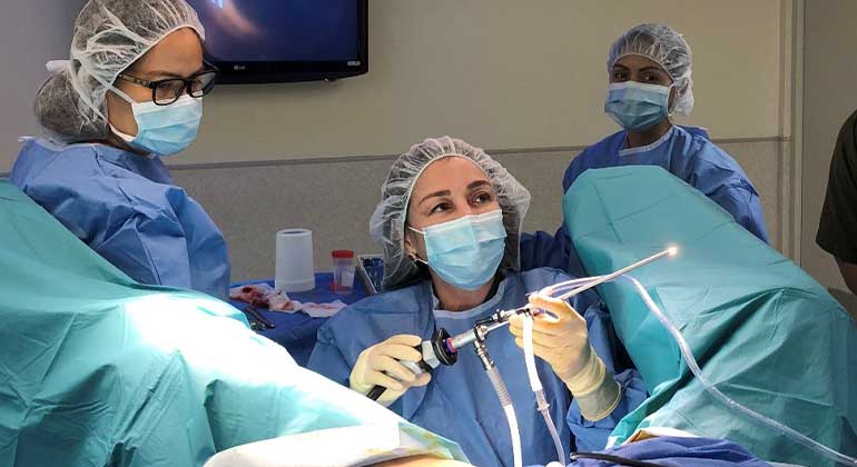
Diagnostic hysteroscopy allows the physician to check the size, shape and lining of a woman’s uterus to diagnose any abnormalities that may be affecting fertility or causing other gynecologic disorders. With the patient under anaesthesia, a thin viewing tool called a hysteroscope is inserted into the vagina and gently moved through the cervix into the uterus, where a liquid solution or carbon dioxide is inserted through the hysteroscope to expand the uterus. Once the uterus is expanded, light and camera on the hysteroscope allow the doctor to see the endometrium (lining of the uterus), ovaries and fallopian tubes on a video screen.
Operative hysteroscopy
Operative hysteroscopy may be performed to correct an abnormal condition found during diagnostic hysteroscopy. Small instruments can be inserted through the hysteroscope to correct problems such as uterine septum, uterine polyps and fibroids, or adhesions. Hysteroscopy is typically an outpatient procedure. If a doctor has any concerns, such as a patient’s reaction to anaesthesia, an overnight hospital stay may be required.


